
Package
Musculoskeletal (MSK) Ultrasound
Detailed ultrasound imaging of joints, tendons, ligaments, and soft tissues to investigate pain, injury, swelling, or restricted movement. Scans are region-specific and ideal for sports-related conditions and general MSK assessment.

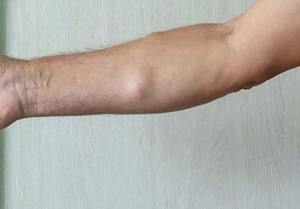
- A detailed assessment of the major muscle groups, including the shoulder, biceps, triceps, and deltoid. We screen for muscle tears, haematomas (internal bruising), and soft-tissue masses. This scan is highly effective for identifying the extent of injury following trauma or sudden strain.
- £180
- Ideal for: Shoulder, Upper arm pain, swelling, suspected muscle tears, or trauma follow-up.
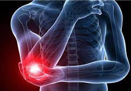
- Evaluation of the common extensor and flexor tendons—often the source of "Tennis" or "Golfer’s" elbow. This scan also assesses the collateral ligaments, the olecranon bursa, and the ulnar nerve for signs of subluxation or entrapment.
- £180
- Ideal for: Elbow pain, swelling, and sports-related injuries
- A focused evaluation of the flexor and extensor muscle groups, tendons, and surrounding soft tissues. We screen for overuse injuries, tenosynovitis, and signs of nerve irritation or entrapment. This assessment provides clarity for patients experiencing persistent pain between the elbow and the wrist.
- £180
- Ideal for: Forearm pain, repetitive strain (overuse) injuries, or suspected nerve-related symptoms.
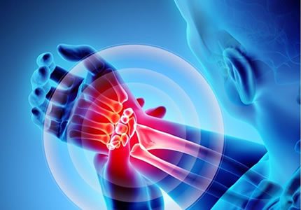
- A focused assessment of the wrist tendons, ligaments, and the carpal tunnel. This scan identifies tenosynovitis, ganglion cysts, and signs of median nerve compression. It is also an essential tool for detecting early osteoarthritic changes or inflammatory joint swelling within the wrist.
- £180
- Ideal for: Wrist pain, tingling or numbness (Carpal Tunnel), and restricted movement.
- A detailed evaluation of the tendons, pulleys, and small joints of the hand and fingers. This scan is designed to identify "Trigger Finger," pulley injuries, and palpable lumps such as small ganglia. We also assess for synovitis (inflammation) and degenerative changes in the finger joints.
- £180
- Ideal for: Finger locking, localised swelling, pain, or palpable lumps in the hand.

- Evaluation of the hip joint, adductor tendons, and the trochanteric bursa. This scan is highly effective at identifying the source of "snapping" hip sensations, bursitis, or proximal muscle tears in the thigh and groin region.
- £180
- Ideal for: Chronic hip pain, stiffness, groin discomfort, or clicking sensations.
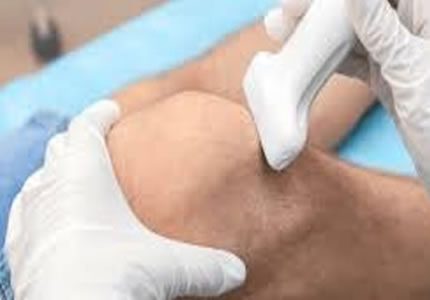
- A detailed assessment of the quadriceps and patellar tendons, collateral ligaments, and joint recesses. We screen for "Baker’s Cysts," joint effusions (fluid), and signs of inflammatory arthropathy to help assess stability and mobility.
- £180
- Ideal for: Swelling, instability, reduced mobility, or post-injury evaluation.
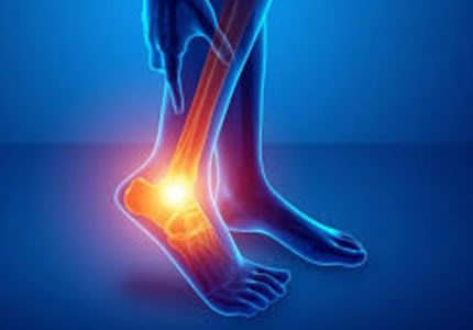
- A comprehensive evaluation of the ankle tendons (including the peroneal and tibial groups), collateral ligaments, and joint spaces. This scan is the clinical standard for identifying sprains, ligament tears, and inflammatory joint changes. It is particularly effective for assessing instability following a sports injury or sudden trauma.
- £180
- Ideal for: Ankle pain, sports injuries, instability, or localised swelling.
- A detailed assessment of the plantar fascia, metatarsal region, and the small tendons and joints of the foot. We screen for common causes of walking-related discomfort, such as Plantar Fasciitis, Morton’s Neuroma, and bursitis. This scan also identifies ganglion cysts or stress-related soft tissue changes in the forefoot.
- £180
- Ideal for: Sharp pain when walking, forefoot symptoms, heel pain, or palpable lumps.
- A focused evaluation for tendinopathy, partial tears, or full-thickness rupture. We assess the tendon structure, the retrocalcaneal bursa, and Kager’s fat pad to provide a clear picture for your physiotherapy or surgical plan.
- £180
- Ideal for: Heel pain, tendon thickening, or suspected rupture.
Package
Lumps, Bumps & Hernia Assessment (Soft Tissue)
High-resolution ultrasound assessment of superficial swellings, subcutaneous masses, and abdominal wall hernias—used for diagnosis, monitoring, or pre-referral imaging.
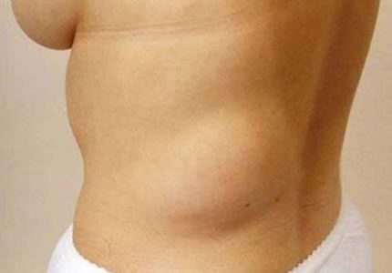
- High-resolution imaging of superficial and subcutaneous masses. We characterise common findings such as lipomas (fatty lumps), epidermoid cysts, ganglia, and vascular lesions. This scan helps distinguish benign findings from those requiring onward referral or fine-needle aspiration.
- £180
- Ideal for: New or persistent swellings, painful lumps, or pre-referral evaluation.
- A dynamic assessment of the abdominal wall and groin. Using Valsalva (coughing) techniques, we identify inguinal, femoral, umbilical, and incisional hernias. We assess the hernia contents and their "reducibility" to support your surgical planning.
- £180
- Ideal for: Intermittent bulges, groin pain, or abdominal wall swelling.
- A dedicated survey designed for the early detection and monitoring of inflammatory conditions such as Rheumatoid or Psoriatic Arthritis. ✧ Inflammatory Joint Survey This specialist package evaluates both hands and wrists for synovitis, tenosynovitis, and early structural erosions. We use Power Doppler to assess active inflammation, helping to differentiate between degenerative wear and active inflammatory patterns. This is an essential tool for monitoring treatment response or reaching an early diagnosis.
- £400
- Ideal for: Suspected rheumatoid arthritis, morning stiffness, or monitoring treatment.